Mesothelium Histology : Webpathology Com A Collection Of Surgical Pathology Images / The fibrous pericardium is the outer layer, and the serous pericardium is the inner layer.the space between the two layers is the pericardial cavity.
Mesothelium Histology : Webpathology Com A Collection Of Surgical Pathology Images / The fibrous pericardium is the outer layer, and the serous pericardium is the inner layer.the space between the two layers is the pericardial cavity.. Serous membranes consist of a single layer of epithelium, named mesothelium, attached to a supporting layer of connective tissue, with a small layer in between, the basal membrane (fig 1). Meaning tissue or columns, and "logia," The inner surface is lined by the stratified squamous epithelium. The connective tissue of the visceral pleura is contiguous with the connective tissue of the pulmonary lobular septae that course through the pulmonary. Neet zoology biomolecules questions &
Results and discussion formation of the lung mesothelium. The thick portions have an histology characteristic of either proximal or distal tubule. The pleura (thoracic cavity), peritoneum (abdominal cavity including the mesentery) and pericardium (heart sac). If the endoscope can go there without puncturing the skin or mucosal membranes and "drawing blood", the surface is covered by epithelium. Each lung projects into an internal body cavity, the pleural cavity, and is covered by a serous membrane (serosa), called visceral pleura.
Multicystic Peritoneal Mesothelioma A Systematic Review Of The Literature from www.degruyter.com When covered by mesothelium, the adventitia is called the serosa. Working in a histology lab means that i get to see a lot of what our body looks like under the microscope. Adrian rad bsc (hons) • reviewer: The connective tissue of the visceral pleura is contiguous with the connective tissue of the pulmonary lobular septae that course through the pulmonary. Red blood cells are also bright red. Epithelial tissues cover all external and internal surfaces of the body. mesothelium, or the tissue formed by mesothelial cells, helps protect the organs by producing a lubricating fluid that allows the organs to move without irritating nerves. The deepest portions of the medulla have only thin segments and collecting ducts.
Simple squamous epithelium flaened squamous cells in a single layer duct linings o1en have simple cuboidal epithelium, like this smallish duct in the pancreas.
The fibrous pericardium is the outer layer, and the serous pericardium is the inner layer.the space between the two layers is the pericardial cavity. histology for pathology cardiac system theresa kristopaitis, md associate professor director of mechanisms of human disease kelli a. In the esophageal mucosa, the epithelium is stratified squamous and the muscularis mucosae is unusually thick. Before to start i would like to provide the summary of this organ. It is not so well defined in smaller lobules (lobuli) as is the case in mammals (especially pigs). histology of a serous membrane. There are many arrangements of epithelial cells such as squamous, cuboidal, and columnar that organize as simple, stratified, pseudostratified, and transitional. The connective tissue of the visceral pleura is contiguous with the connective tissue of the pulmonary lobular septae that course through the pulmonary. Move mouse coursor upon an image and you can see the image without labells. Simple columnar epithelium (absorptive cells with brush border, terminal bars, goblet cells), basement membrane: Avascular tissues with closely apposed cells without intervening intercellular substances. Skin is covered by epithelium (that's the epidermis layer. Adrian rad bsc (hons) • reviewer:
By releasing a lubricating fluid, the mesothelium allows the organs to move more freely within the body cavity; histology for pathology cardiac system theresa kristopaitis, md associate professor director of mechanisms of human disease kelli a. Villous processes of varied architectural complexity and surface epithelial crypts were found in the ovary but not in t … mesothelium is a simple squamous epithelial tissue which forms the surface of the serosa in the major body cavities (peritoneal, pleural, and pericardial). Refer to the diagram at the end of this chapter for the tissue orientation and consult.
Mesothelium An Overview Sciencedirect Topics from ars.els-cdn.com The morphology of the ovarian mesothelium (surface epithelium) and of other pelvic and extrapelvic mesothelia was investigated in the rabbit by use of scanning electron microscopy. histology is the microscopic counterpart to gross anatomy, which looks at larger structures visible without a microscope. Distributed unequally among different types of ct.collagen and elastin are the 2 major proteins that make up ct. Comes from the greek words "histos," Working in a histology lab means that i get to see a lot of what our body looks like under the microscope. Red blood cells are also bright red. histology for pathology cardiac system theresa kristopaitis, md associate professor director of mechanisms of human disease kelli a. The surface of the liver is covered by a peritoneal layer of mesothelium and a very thin glisson's capsule.
If the endoscope can go there without puncturing the skin or mucosal membranes and "drawing blood", the surface is covered by epithelium.
It attached to the lower surface of the liver. The organ of focus this quarter is the fallopian tube. Before to start i would like to provide the summary of this organ. The pleura (thoracic cavity), peritoneum (abdominal cavity including the mesentery) and pericardium (heart sac). Regional histology of the digestive tract. You should read this full article to know about the liver histology.this is the ultimate guide to learn normal liver histology in details. In slide 29 and slide 176, this type of epithelium lines the luminal (mucosal) surface of the small and large intestines, respectively. Which type of cartilage forms the skeleton of the fetus? 500 practice questions for histology 1. Vessels, adipocytes and an outer layer of mesothelium mesothelium secretes pericardial fluid covers and protects the heart cardiac valves 4 valves 2 av (mitral and tricuspid) in the chambers. This diagram shows that the simple squamous epithelium of the tunica adventitia layer of the heart (mesothelium) is also the visceral layer of the serous pericardium. Quarterly i will share with you some of my photos from the microscopic world of our inner space and tell you a little bit about what we're looking at. histology of a serous membrane.
500 practice questions for histology 1. Serous membranes consist of a single layer of epithelium, named mesothelium, attached to a supporting layer of connective tissue, with a small layer in between, the basal membrane (fig 1). Mucosa, submucosa, muscularis externa, and adventitia. Each lung projects into an internal body cavity, the pleural cavity, and is covered by a serous membrane (serosa), called visceral pleura. Uruj zehra mbbs, mphil, phd last reviewed:
Mesothelium High Resolution Stock Photography And Images Alamy from c8.alamy.com Malignant mesothelioma frequently involves severe respiratory problems. A tissue is a group of cells, all of the same type, working together to perform a function. The virus sv40 may also be a factor in the disease in some people. The connective tissue of the visceral pleura is contiguous with the connective tissue of the pulmonary lobular septae that course through the pulmonary. Mesenteries , the sheets of connective tissue which bind together the loops of the gi tract, have the same composition as serosa and, like the serosa , and are covered on exposed faces by. The pleura (thoracic cavity), peritoneum (abdominal cavity including the mesentery) and pericardium (heart sac). histology is defined as the scientific study of the microscopic structure (microanatomy) of cells and tissues. If the endoscope can go there without puncturing the skin or mucosal membranes and "drawing blood", the surface is covered by epithelium.
All of the above e.
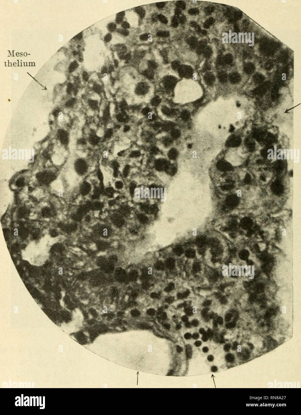
It attached to the lower surface of the liver. mesothelium (simple squamous epithelium) view from surface stained with silver nitrate. Which means study.the word "histology" This slide shows mesothelium, simple squamous epithelium on the outside of an organ.simple squamous epithelium is: Epithelial cells are the cellular components of the epithelium. This diagram shows that the simple squamous epithelium of the tunica adventitia layer of the heart (mesothelium) is also the visceral layer of the serous pericardium. Embryologically it develops from the foregut and it spans the upper right and part of left abdominal quadrants. The mesothelium is a membrane that forms the lining of several body cavities: (really a modified mesothelium.) slide 2 cortex of ovary. The fibrous pericardium is the outer layer, and the serous pericardium is the inner layer.the space between the two layers is the pericardial cavity. Results and discussion formation of the lung mesothelium. 500 practice questions for histology 1. All of the above e.
mesothelium, or the tissue formed by mesothelial cells, helps protect the organs by producing a lubricating fluid that allows the organs to move without irritating nerves mesothelium. Somewhat deeper lie several small, primary (primordial) follicles.


0 Comments